
Parts of a Compound Microscope Labeled (with diagrams) Medical Pictures and Images (2023
Labeling the Parts of the Microscope This activity has been designed for use in homes and schools. Each microscope layout (both blank and the version with answers) are available as PDF downloads. You can view a more in-depth review of each part of the microscope here. Download the Label the Parts of the Microscope PDF printable version here.

Microscope Diagram Labeled, Unlabeled and Blank Parts of a Microscope
A Study of the Microscope and its Functions With a Labeled Diagram To better understand the structure and function of a microscope, we need to take a look at the labeled microscope diagrams of the compound and electron microscope. These diagrams clearly explain the functioning of the microscopes along with their respective parts.
16 Parts of a Compound Microscope Diagrams and Video Microscope Clarity
Simple microscope is a magnification apparatus that uses a combination of double convex lens to form an enlarged, erect image of a specimen. The working principle of a simple microscope is that when a lens is held close to the eye, a virtual, magnified and erect image of a specimen is formed at the least possible distance from which a human eye.

Microscope diagram Tom Butler Technical Drawing and Illustration Projects Pinterest
The hand magnifying glass can magnify about 3 to 20×. Single-lensed simple microscopes can magnify up to 300×—and are capable of revealing bacteria —while compound microscopes can magnify up to 2,000×. A simple microscope can resolve below 1 micrometre (μm; one millionth of a metre); a compound microscope can resolve down to about 0.2 μm.
Cells and Microscopes
A light microscope is a biology laboratory instrument or tool, that uses visible light to detect and magnify very small objects and enlarge them. They use lenses to focus light on the specimen, magnifying it thus producing an image. The specimen is normally placed close to the microscopic lens.
How to Use a Microscope
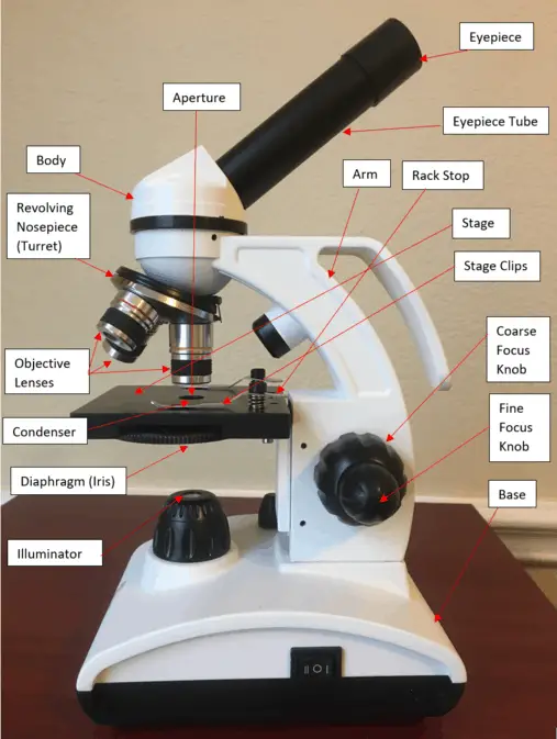
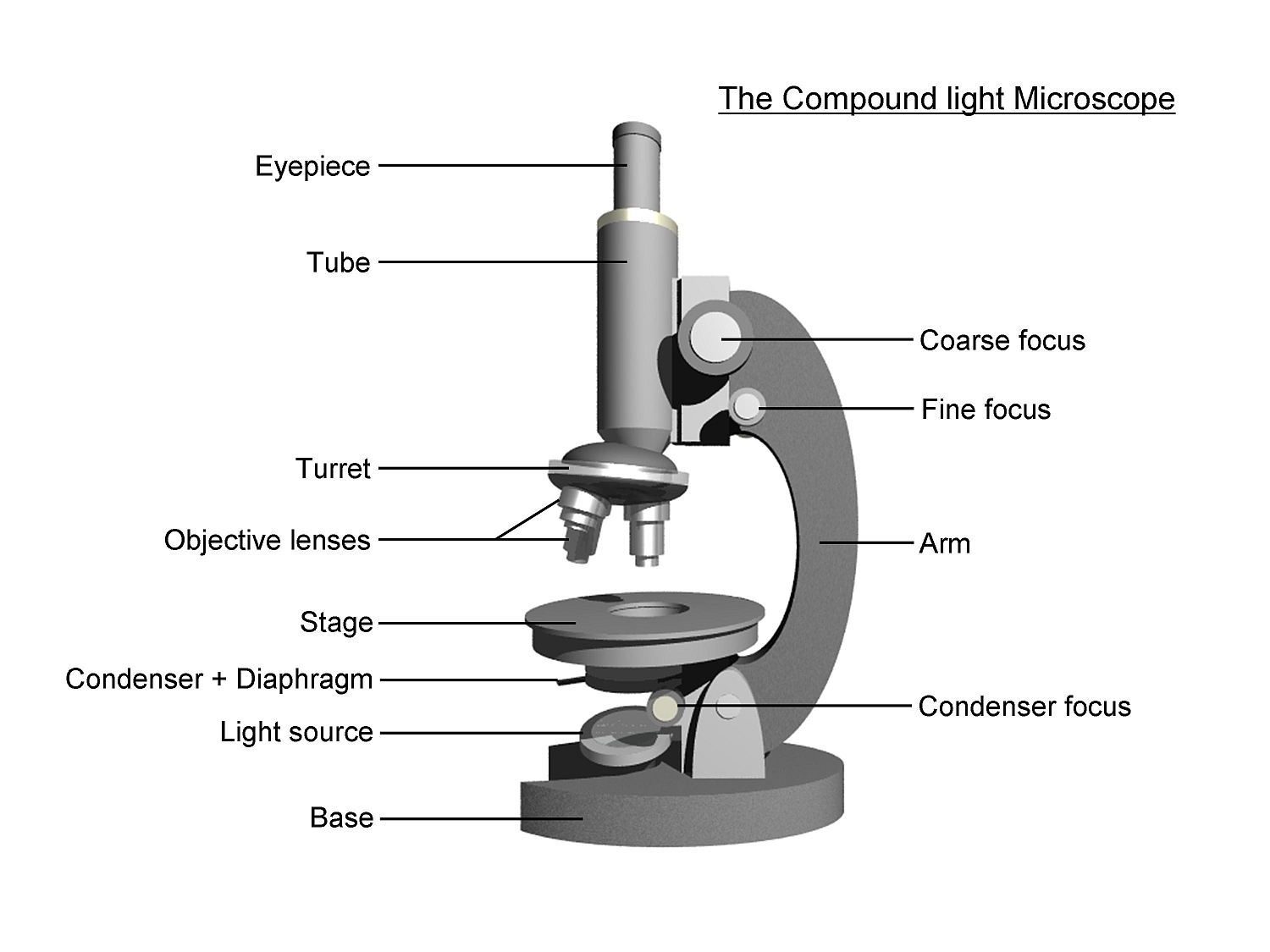
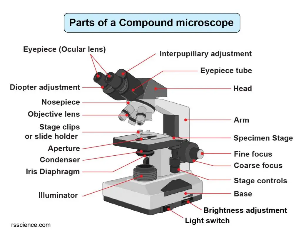
Labeled diagram of a compound microscope Major structural parts of a compound microscope Optical components of a compound microscope Eyepiece Eyepiece tube Objective lenses Nosepiece Specimen stage Coarse and fine focus knobs Rack stop Illuminator Condenser Abbe condenser Iris Diaphragm Condenser Focus Knob Summary An overview of microscopes
Parts of a Compound Microscope — Learning in Hand with Tony Vincent
What type of labels should I use for my microscope? Properly labeling your microscope is an important part of using it efficiently and effectively. Choosing the right type of label for your microscope is essential to ensure that it remains legible and lasts for the entire lifetime of the microscope.
Parts Parts And Functions Of A Microscope
Microscopes help forensic professionals in examining hair, fragments, skin cells, and evidence material to determine the exact cause of an event and help the law enforcement and investigating agencies in nabbing the culprits and bringing them to justice.
Ag Biology Unit 2
A compound microscope is used for viewing small samples or pieces of a larger specimen at higher magnification. This type of microscope uses transmitted light, where the light must pass through the specimen to view it. Figure 9.1.2 9.1. 2: A labeled compound microscope, with the stage for the specimen located between the lenses and the light.

Labeled Microscope Diagram Tim's Printables
The labeling worksheet could be used as a quiz or as part of direct instruction. Students label the microscope as you go over what each part is used for. Then they answer short fill in the blank sentences about the proper use of the microscope. The google slides shown below have the same microscope image with the labels for students to copy.
Compound Microscope Parts Labeled Diagram and their Functions (2023)
A microscope is an instrument that magnifies objects otherwise too small to be seen, producing an image in which the object appears larger. Most photographs of cells are taken using a microscope, and these pictures can also be called micrographs. From the definition above, it might sound like a microscope is just a kind of magnifying glass.
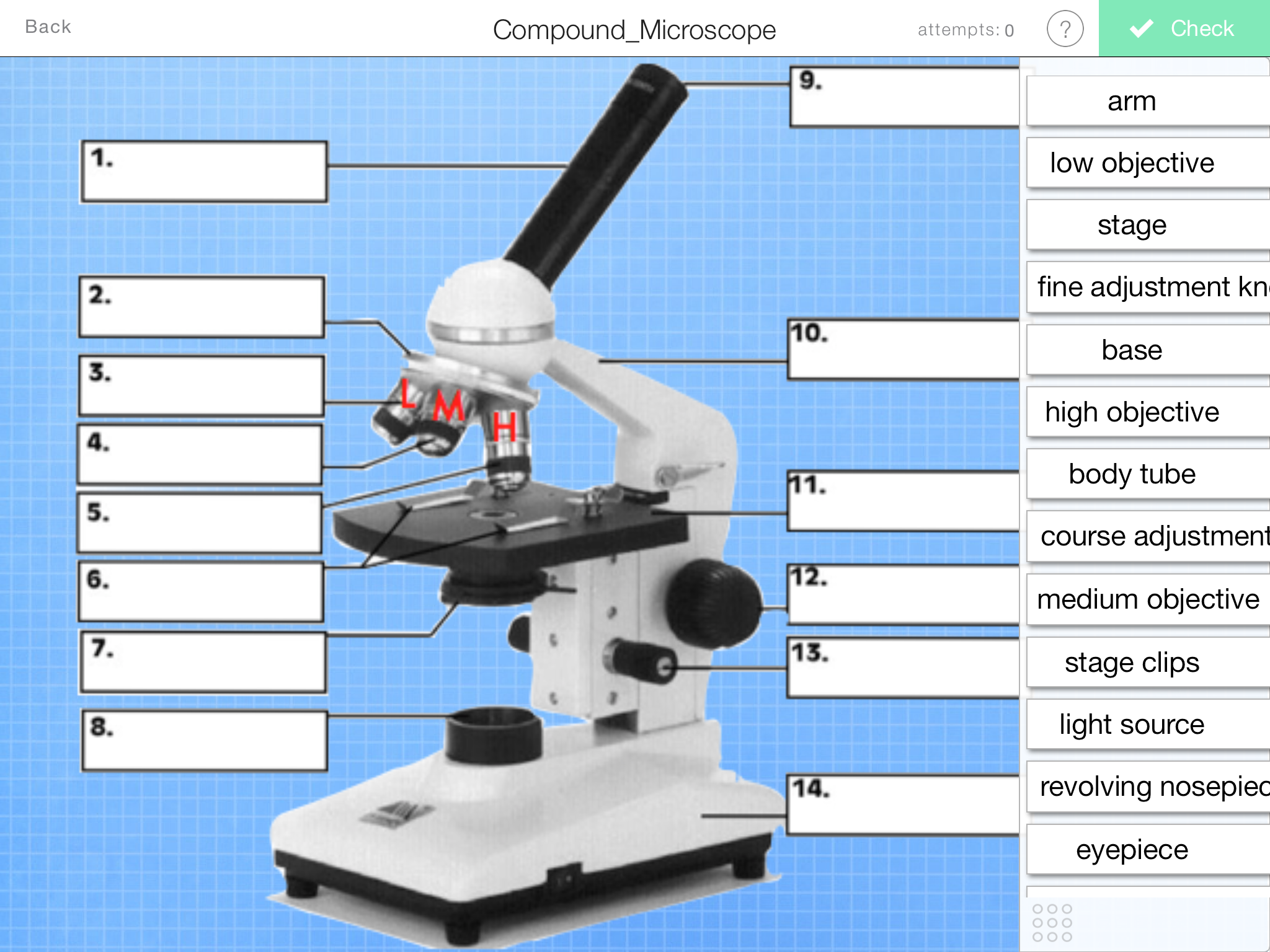
Parts of a Microscope Labeling Activity
Microscopes are instruments that are used in science laboratories to visualize very minute objects, such as cells and microorganisms, giving a contrasting image that is magnified. Microscopes are made up of lenses for magnification, each with its own magnification powers.

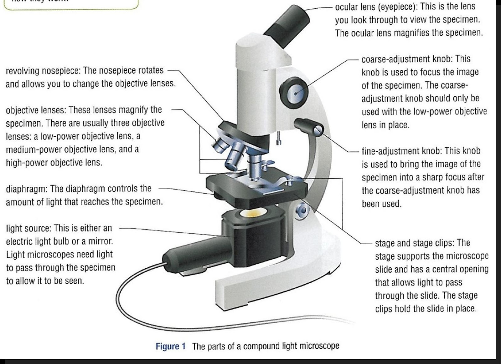
Parts of a microscope with functions and labeled diagram
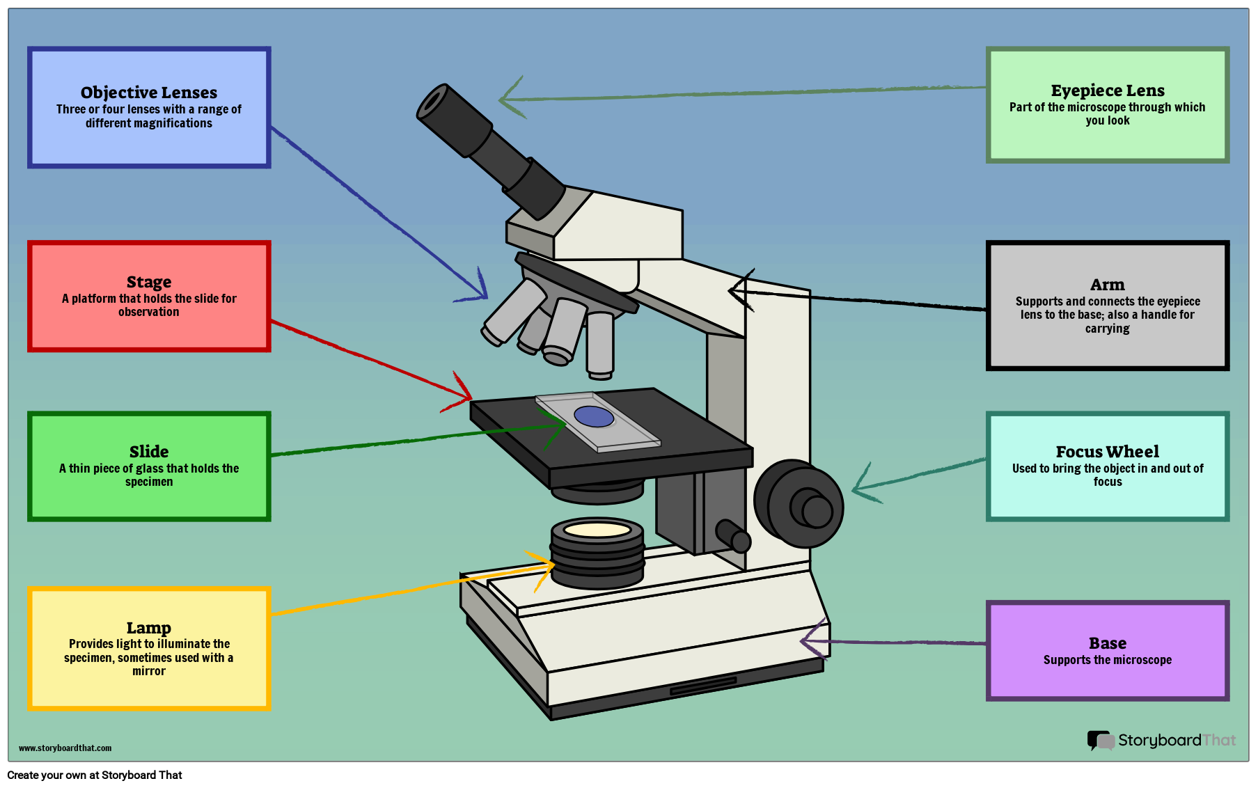
Create a poster that labels the parts of a microscope and includes descriptions of what each part does. Click "Start Assignment". Use a landscape poster layout (large or small). Search for a diagram of a microscope. Using arrows and textables label each part of the microscope and describe its function. More options.

Simple Microscope Definition, Principle, Magnification, Parts, Applications
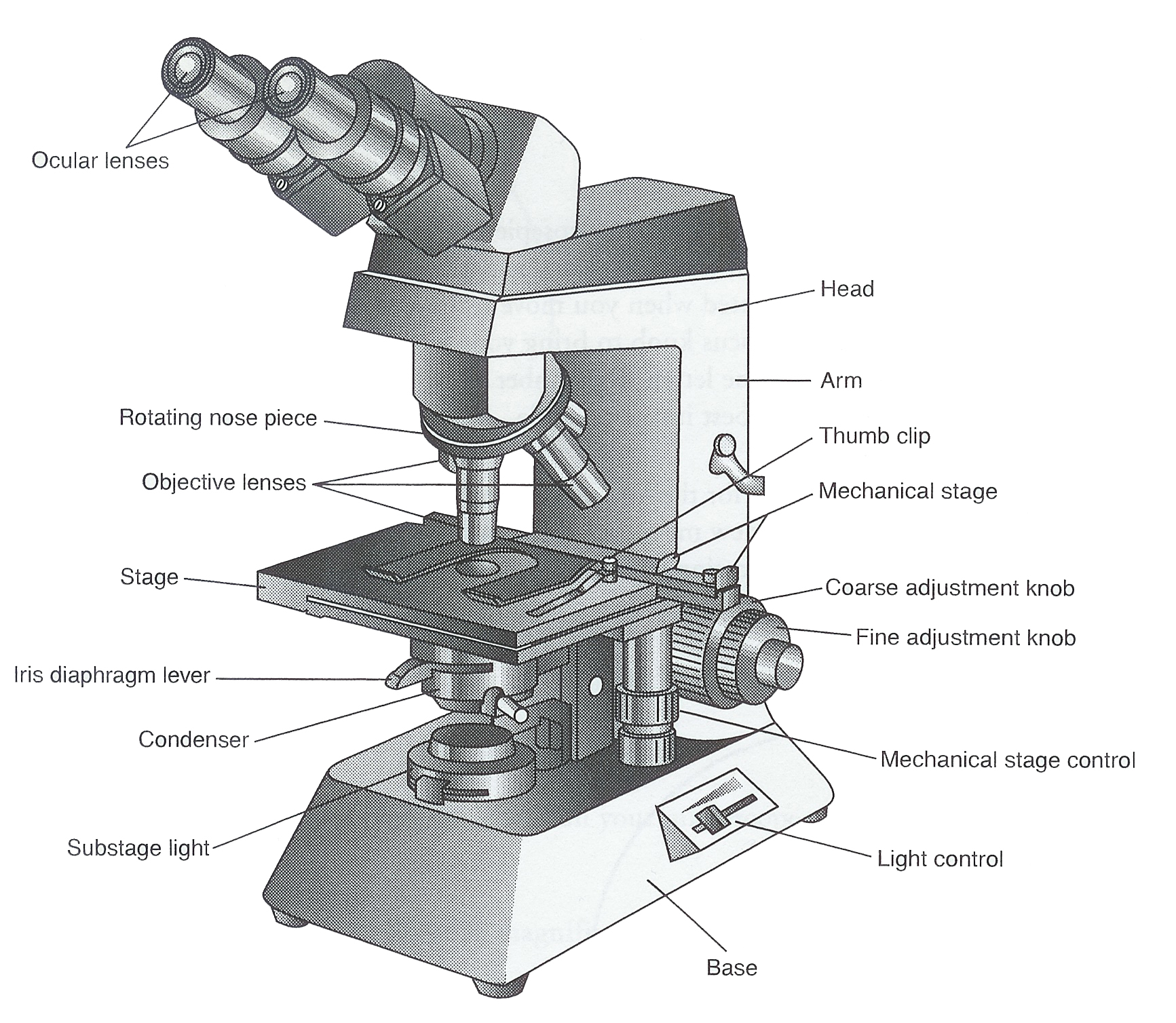
Parts of Compound Microscope (Labeled Pictures) a. Mechanical Parts of a Compound Microscope Foot or Base Pillar Arm Stage Inclination Joint Clips Diaphragm Nose piece/Revolving Nosepiece/Turret Body Tube Adjustment Knobs b. Optical Parts of a Compound Microscope Eyepiece lens or Ocular Mirror Objective Lenses
The Parts of a Compound Microscope and How To Handle Them Correctly Human Anatomy and
It also allows the specimen to be labeled, transported, and stored without damage. Stage: The flat platform where the slide is placed. Stage clips: Metal clips that hold the slide in place. Stage height adjustment (Stage Control): These knobs move the stage left and right or up and down.
Dissecting Microscope Uses New York Microscope Company
Label the microscope Interactive Add to collection Use this interactive to identify and label the main parts of a microscope. Drag and drop the text labels onto the microscope diagram. base high-power objective eye piece lens coarse focus adjustment diaphragm or iris stage fine focus adjustment light source Download Exercise Tweet